Table of Contents
Serratus Anterior Anatomy
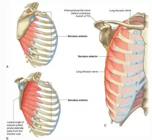
Serratus anterior (also known as the boxers muscle) is a thick and broad muscle of the thorax with a strong and well-developed fascia that covers it. This muscle covers the lateral part of the thorax and also forms the medial wall of the axilla. This muscle was named Serratus (in Latin means a Saw) because of its saw-toothed appearance of its fleshy slips or digitations. The muscular slips pass posteriorly and then medially to attach to the whole length of the anterior surface of the medial border of the scapula, including its inferior angle.
Serratus Anterior Action and Function
- The Serratus anterior is a powerful protraction muscle of the pectoral girdle that is used when punching or reaching anteriorly, hence it is called boxer’s muscle.
- The strong lower part of Serratus anterior rotates the scapula and elevates the glenoid cavity in order for the arm to be raised above the shoulder.
- Serratus anterior also supports the scapula by stabilizing it and making it closely applied to the thoracic wall so that other muscles can use the scapula for movements of the humerus.
- The Serratus anterior also holds the scapula against the thoracic wall during push-ups or when pushing against resistance such as when pushing a car.
Serratus Anterior Origin and Insertion
The origin of Serratus anterior is from a series of digitations from the first eight ribs. The first digitation arises from the first and second ribs with fibers of C5 cervical segment whereas other digitations arise from their corresponding ribs (the 3rd digitation arising from the 3rd rib, 4th digit of origin from 4th rib and so on till it reaches the 8th digitation that arises from the 8th rib); the 3rd and 4th digitations are innervated by C6 cervical segment of the spinal cord while the lower four digits join with that of the external oblique muscle and are supplied by the C7 segments. Passing over the upper border of serratus anterior is a neurovascular bundle from the posterior triangle to the axilla.
The Serratus anterior muscle then inserts on the inner and costal surface of the scapula with the first and second digitations inserting at the superior angle of the scapula, the third and fourth digitations inserting as a thin sheet to the length of the vertebral border and the lowest four at the inferior angle.
Serratus Anterior Nerve Supply
Serratus anterior is innervated by the Long thoracic nerve (also known as the nerve to serratus anterior) which arises from the roots of the brachial plexus (C5, C6, C7). The branches from C5 and C6 unite in the scalenus medius muscle and emerge from its lateral border as a single trunk which enters the axilla by passing over the first digitation of serratus anterior. The contribution from C7 also passes over the first digitation of serratus anterior. The long thoracic nerve lies behind the mid axillary line, behind the lateral branches of the intercostal arteries on the muscles surface but deep to the fascia, and therefore is protected in surgical operations on the axilla.
Serratus Anterior Blood Supply
The blood supply of serratus anterior muscle is from the lateral thoracic artery which also supplies other structures on the lateral aspect of the breast and thorax (chest). Other arterial supply to the serratus anterior include the Superior thoracic artery (a branch of the axillary artery) this supplies the upper part of the serratus anterior while the lower part is supplied by the thoracodorsal artery (a branch of the subscapular artery).
Tests for Serratus Anterior
To test the serratus anterior (or the function of the long thoracic nerve that supplies it), the hand of the outstretched limb is pushed against a wall. If the Serratus anterior muscle is acting normally, several digitations of the muscle can be seen and palpated.
Serratus Anterior Injury and Paralysis
When the serratus anterior is paralyzed because of injury to the long thoracic nerve, it causes displacement of the scapula the medial border of the scapula moves laterally and posteriorly away from the thoracic wall giving the appearance of a winged scapula. When the arm is raised, the medial border and inferior angle of the scapula pull markedly away from the posterior thoracic wall, a deformation known as a winged scapula. The arm also cannot be abducted above the horizontal position because the serratus anterior is unable to rotate the glenoid cavity superiorly to allow complete abduction of the limb.
